Part 1
What is feline asthma?
‘Feline asthma’, sometimes referred to as ‘chronic bronchitis’ or ‘allergic airway disease’, is a term used to describe chronic lower airway disease affecting the small tubes (bronchioles) within the lungs of cats.
In human medicine, asthma and chronic bronchitis are considered two independent conditions; however, some people may suffer from both conditions. At this stage, it is assumed that most cats who develop signs of lower airway disease have a similar condition to humans. The exact cause (and mechanism) as to why certain cats develop this syndrome is still unknown.
Asthma is a condition that results from exposure to airway triggers (an irritant or allergen) which provokes an inflammatory response. This inflammatory response results in narrowing of the airway tubes due to spasm/contraction of the surrounding smooth muscle, and excessive mucous production. In combination, these changes can lead to the inability to draw a deep breath, exercise intolerance, panting, coughing, and high-pitched whistling sounds (wheezing). Sometimes a chronic cough is the only sign noticed by cat owners. However, an acute asthmatic crisis can occur at any time and present itself as a life-threatening event. Asthmatic airway constriction can happen spontaneously or as a type of allergic reaction.

Figure 1: Asthma can be more common in Siamese and Oriental breeds
Which cats are likely to develop feline asthma?
It is believed that any breed, age, or sex can develop clinical signs relating to asthma. Most cats diagnosed with this condition are typically middle aged adults (5-9 years old) but it can start in both young and old cats. There also appears to be a strong breed association amongst Siamese/Oriental breeds and this may suggest an inherited link to this condition.
How is the diagnosis of feline asthma made?
A step toward making a diagnosis of feline asthma is for your veterinary surgeon to take x-rays of your cat’s chest (this is done whilst they are awake and not under a general anaesthetic). Because many cats will present as an emergency, your veterinary surgeon will most often need to stabilise your cat first with as little restraint as possible in an ‘oxygen-rich’ environment (this is a purpose built ‘tent’ or cage containing high levels of oxygen). Because of the constricted airways, the actual volume of air an asthmatic patient can move in and out of the lungs with each breath is markedly reduced. Because of this, your cat is working extremely hard to inhale and exhale air from the lungs. The abdomen may appear expanded as it attempts to push air out. Some cats will also gulp air and have a swollen stomach. The breaths are often short and shallow, and your cat may even be breathing with their mouth open in an effort to move the largest possible amount of air in and out of the lungs.

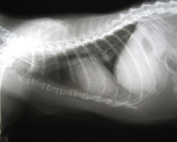
Figure 2: Chest x-rays can help rule out many diseases responsible for changes in breathing pattern such as fluid on the lungs (‘pulmonary oedema’), fluid around the lungs (‘pleural effusion’), masses (abscesses or cancer), ruptured diaphragm, and patchy lung patterns commonly found with pneumonia.
Once your cat is considered to be clinically stable in a calm and oxygen-rich environment, and they are tolerant to handling, then conscious chest x-rays (ie. with your cat awake and not anaesthetised) can be taken to rule out other causes such as fluid on the lungs (pulmonary oedema) and fluid around the lungs (pleural effusion) associated with congestive heart failure.
Classically, x-rays from asthmatic cats will show areas of what we call ‘air-trapping’. Air-trapping simply means that the small airways have constricted so much that inhaled air cannot be fully exhaled from the lungs. Asthmatic lungs will also appear larger than normal and the diaphragm may seem flattened due to over-inflation.
Inflammation and mucus build up within the airways will cause the airway walls to appear thickened in the x-ray. Your veterinary surgeon may use specific terms to describe ‘classic’ signs of airway thickening as either ‘doughnuts’ (when viewing the airway end-on) and/or ‘tramlines’ (when viewing the airway from the side).
Other diseases affecting the airways (inhaled foreign body, fluid (pleural effusion), blood (haemothorax), air (pneumothorax) or pus surrounding the lungs (pyothorax), bacterial/viral/parasitic pneumonia, heart disease, and cancer) can present in similar ways to asthma and require further investigations, in addition to conscious chest x-rays, to rule out these other potential underlying causes.
Further tests may include:
- Blood tests
- Faecal tests (looking for lungworm eggs/larvae)
- ‘Inflated’ chest x-rays under general anaesthetic to assess the lungs, diaphragm, space surrounding the lungs (pleural space) and heart
- Bronchoscopy: this involves passing a long flexible camera into the lungs and assessing for any abnormalities such as foreign bodies or areas of pus coming from a specific area of the lung
- Lower airway washes (Bronchioalveolar lavage or ‘BALs’): these are often performed under general anaesthesia, with or without the use of a bronchoscope, to collect samples for the pathologist to examine under a microscope. Small volumes of sterile saline are flushed into the lungs and immediately aspirated back into the syringe. These samples are sent for a variety of tests and to look for abnormal inflammatory cell numbers, bacteria, parasites, and/or cancer cells as a cause to the clinical signs

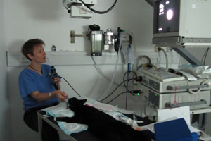
Figure 3: Endoscopy of the airways can be performed under general anaesthetic. This can help your veterinary surgeon to assess the structure and function of the larynx, or voicebox (‘laryngoscopy’), the trachea, or windpipe (‘tracheoscopy’), and the smaller airways leading to the lungs (‘bronchoscopy’). This will also allow your vet to collect airway samples which can be evaluated for signs of parasites, infection, or cancer.
Lung washes from the lower respiratory tract from cats with clinical signs suggestive of asthma or chronic bronchitis, but with normal chest x-rays, may be helpful in some cases in achieving a diagnosis. Types of inflammatory white cells called ‘eosinophils’ and ‘neutrophils’ are often abundant in the secretions of asthmatic/chronic bronchitis patients; however, interpretation of this finding is not ‘black and white’ as these cells can also occur in normal feline respiratory secretions.

Figure 4: This is an example of a lung wash (‘bronchioalveolar lavage’ or ‘BAL’) from a cat with a chronic cough. This cat was diagnosed with mycoplasma pneumonia and secondary chronic bronchitis. A normal lung wash will appear clear like water with very little foam on top. This sample has a large amount of mucous, and thick, turbid, frothly material.
Can the response to therapy be used as a diagnostic test?
Sometimes diagnostic tests leave room for question and your veterinary surgeon has to simply go with medical treatment for presumptive asthma/chronic bronchitis and regard response to therapy as evidence for a correct diagnosis.
Currently in practice, both asthma (reversible bronchoconstriction) and chronic bronchitis are treated in very similar ways. Generally speaking, managing the secondary infections (e.g Mycoplasma, Bordetella bronchiceptica, lung worm) should be identified and treated and any exacerbating causes should be eliminated from the living environment. This includes identifying and eliminating potential inhaled household irritants/allergens such as dusty litter, smoking, and aerosol sprays.
In the second part of this article, Elise Robertson explains the treatment options available for cats with asthma.
 The Veterinary Expert| Pet Health
The veterinary expert provides information about important conditions of dogs and cats such as arthrits, hip dysplasia, cruciate disease, diabetes, epilepsy and fits.
 The Veterinary Expert| Pet Health
The veterinary expert provides information about important conditions of dogs and cats such as arthrits, hip dysplasia, cruciate disease, diabetes, epilepsy and fits.